Discover more about these symptoms and next steps

Choose the option below that sounds most like you to discover your best solution

Eye treatment options can be confusing. We’ve made the journey as straightforward as can be
Get a quick overview of everything you need to know about gaining visual freedom

Caring for your eyes
Today is World Sight Day. A day to focus the world's attention on the importance [...]

Eye Institute Wellington Opening Evening
Eye Institute Wellington Opening Evening On Friday 19th May Eye Institute Wellington celebrated the opening of our stunning new Wellington [...]
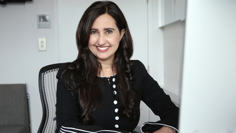
The Ophthalmologist Power List 2023
The Ophthalmologist Power List 2023 Congratulations to Professor Helen Danesh-Meyer for being recognised [...]
Learn more about treating eye floaters and flashes from authoritative sources.
Note: These links will take you off our website